3D Printed Biopatch to Revolutionize Treatment for Heart Attacks

Minnesota university researchers have tested a 3D printed biopatch on mice which can heal scarred tissue after a heart attack.
When it comes to matters of the heart, a team of University of Minnesota biomedical engineering researchers are leading the way. The team is working on a way to heal scarred heart tissue after a heart attack. The process involves a 3D bioprinted patch and could majorly advance heart attack treatment.
Researchers from University of Wisconsin-Madison and University of Alabama-Birmingham made use of 3D bioprinting techniques as well as lasers. Most notably, the team were able to put human heart stem cells on a matrix that began to grow in the lab. Check out the resulting beating patch in the video below.
It’s expected that this research could have a huge advantage for patients worldwide. Heart disease kills more than 360,000 people in the USA every year alone, according to the American Heart Association. It is now the main cause of death in the States.
The reason that heart disease is so deadly is that after a heart attack, the body can’t replace heart muscle cells. This means the patient is then at risk of future heart problems.
Animal Testing the 3D Printed Biopatch
Currently, the researchers are testing the biopatch by using it on mice which are given simulated heart attacks. They found that after just four weeks, the mouse’s heart increased in its functional capacity.
The reason for this is that the 3D printed biopatch is made up of cells from the heart. Therefore, the body can absorb it easily.
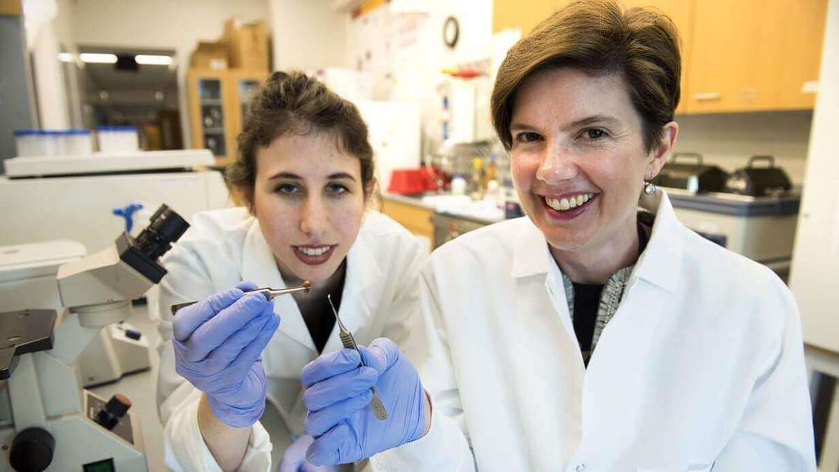
Brenda Ogle, University of Minnesota’s associate professor of biomedical engineering, explains: “We feel that we could scale this up to repair hearts of larger animals and possibly even humans within the next several years.”
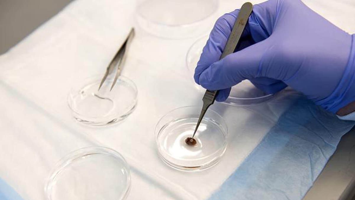
In order to develop the biopatch, the team used a 3D scan of the structural proteins of heart tissue. Then, the researchers 3D print the model with proteins native to the heart.
Since 3D printing allows for one micron resolution, it is the only process that can achieve the required structures. As well as this, it is an especially important feature when mimicking native heart tissue.
“We were quite surprised by how well it worked given the complexity of the heart,” Ogle said. “We were encouraged to see that the cells had aligned in the scaffold and showed a continuous wave of electrical signal that moved across the patch.”
However, the team are now moving on with their research. Next, they intend on testing the 3D printed biopatch on a pig’s heart. The reason for this is that it’s closer to the size of a human’s heart.
Want to find out more? You can read the full research paper on the Circulation Research website.
Source: University of Minnesota
License: The text of "3D Printed Biopatch to Revolutionize Treatment for Heart Attacks" by All3DP is licensed under a Creative Commons Attribution 4.0 International License.