Sara Ryding, a researcher at Deakin University in Australia, had a theory that climate change, over the past century, was affecting the shape of birds’ beaks, which is one way they regulate body temperature. If this were true, it could show how fast species adapt to environmental changes and have profound implications for other species — and us, too.
Proving it meant measuring the beaks of thousands of birds kept in collections at museums across the country dating back to the late 1800s. A daunting task with traditional measuring tape and calipers, but with a hand-held 3D color scanner, Ryding went from theory to proof in about a year.
Her findings that warm-blooded animals are adapting to climate change by shapeshifting some of their features faster than would usually be expected made headlines worldwide.
Enabling Faster, More Accurate Studies
The ability to point a 3D scanner at an object, whether it’s a museum fossil or an ancient relic, and record submillimeter data on length, width, height, surface features, and color is accelerating all types of research.
For example, consider the repository of 3D scan data from museums around the world called MorphoSource, which now has more than 66,000 scans of everything from prehistoric shark teeth to ancient Egyptian water vessels. In 2021 alone, the repository is cited in more than 400 research studies.
For Ryders’ bird research, however, she needed to travel and scan bird specimens on-site.
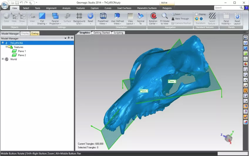
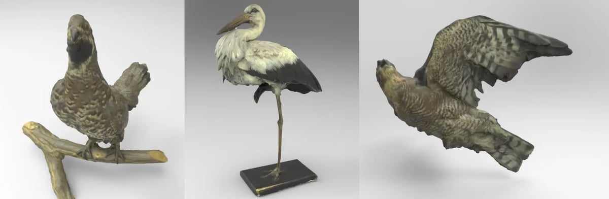
The traditional way of measuring bird beaks is with digital calipers to calculate the length, width, and depth. From there, these dimensions are plugged into an equation that gives researchers the surface area of a cone of the same size. However, considering that bird beaks come in a wide variety of complex shapes, Ryding found that reducing the measurements of a beak down to a simple cone shape would lose key geometry and eliminate important anatomical surface data essential for her study.
“I am focusing on really different birds, from ducks to songbirds, as well as birds of prey,” says Ryding, “…and when you’re studying such diverse species, you’re bound to see really different beak shapes. That’s why I think 3D scanning is much better for this kind of application since you’re capturing all the organic surface anatomy, so nothing gets left out.”
One of the other difficulties of the manual measurement approach is that of operator error. Specifically, this means that the measurements taken can vary from researcher to researcher due to slightly different caliper placement, resulting in significant enough variations to affect the results of the data, even with the best of intentions and experience.
In Ryding’s words, “you can reduce operator error down to a bare minimum since almost every scan is the same, from person to person. This is what every researcher is aiming for. The greater the accuracy of the dataset, the more clearly we can see correlations, which in turn help us reach our conclusions.”
The No-Touch Research Tool
Another clear advantage for 3D scanning in research is to not damage delicate artifacts.
When Monash University researchers Douglass Rovinsky and Justin W. Adams visited museums and university collections around the globe to digitally capture hundreds of specimens of various species needed for their research of the now-extinct Thylacine, they required a non-destructive method of capturing data.
“When we’re given access to a certain specimen at a museum, the last thing the curator wants is to worry about any scratches or other damage from calipers or manual measurement devices, not to mention excessive handling for repositioning,” says Rovinsky. “And placing any markers or targets on these specimens would be unthinkable.”
With very little contact and no risk of damage, in just minutes per specimen, the team scanned a total of 223 crania from 57 species with their Artec Space Spider 3D scanner.

Selecting Scanning Tech
When it came to which 3D scanner was the best fit for the job, Ryding and her team selected the Artec Scan Spider and Scan Studio software with the help of a local equipment distributor.
“The space spider was really beneficial for this work for a number of reasons,” says Ryding. “It handles different sizes of birds and bird beaks quite well, and it’s portable and can be easily taken on flights, which means I was able to fly to museums around the country to collect data. If the scanner hadn’t been portable, the project would not have been doable.”
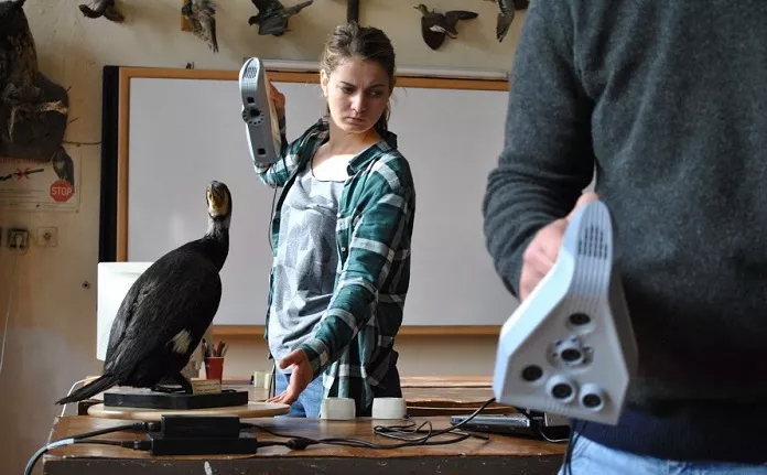
On her scanning days, Ryding goes into a museum with one or more particular species in mind.
With smaller birds, she can scan about 40 or 50 in one afternoon. For larger birds, about 30 to 40 is a more realistic goal.
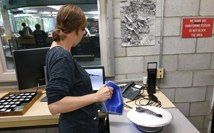
Ryding’s scanning workflow is simple: she places the bird on its back on a turntable. Then, while rotating the turntable slowly, she scans the bird capturing all the essential beak anatomy in a single sweep. Over the course of several months, she scanned more than 3,000 birds with the ultimate target of 6,000 specimens.
Ryding processes the scans in Artec Studio software, either back at home or at her university.
The research is ongoing but so far, it has shown that just over the past century, the beaks of the birds being studied have grown approximately 4% to 10% larger in size.
In regards to broadening the scope of this research well beyond the borders of Australia, Ryding is hopeful that more researchers will add to the existing pool of data, making it possible to accurately quantify and compare the shape-shifting that’s taken place in various species of birds (or other animals) across regions, continents, and hemispheres.
“If we want to facilitate our studies, from researcher to researcher, having directly comparable, accurate data is imperative,” says Ryding. “Manual measurements are no longer sufficient.”
Lead image source: PhD candidate Sara Ryding 3D scanning an Australian galah (Eolophus roseicapilla) with Artec Space Spider (Photo: Sara Ryding)
License: The text of "Ditch the Calipers, Handheld 3D Scanners Accelerate Research" by All3DP Pro is licensed under a Creative Commons Attribution 4.0 International License.