Researchers 3D Print Tissues with Entangled Vascular Networks

Scientists at Rice University developed a new technique for bioprinting tissues, bringing us one step closer to 3D printed organs.
Bioengineers at Rice University developed a new technique which uses bioprinting to create entangled vascular networks to mimic the human body’s natural passageways for vital fluids, including blood and air.
Their research was published on the cover of the May 3 issue of Science.
This breakthrough could bring us closer to bioprinting healthy, functional organs and reduce the need for organ transplants. Every day, 20 people die while waiting for an organ transplant. For those who do receive an organ, there is always a high risk of rejection. By 3D bioprinting a customized organ, this risk would be reduced.
One of the bioengineers that led the research is Jordan Miller, an assistant professor of bioengineering at Rice University’s Brown School of Engineering. He explains that a barrier to building these organs is their complexity.
In order to make them functional, they need a “complex vasculature that can supply nutrients” along with their own vascular networks “like the airways and blood vessels of the lung or the bile ducts and blood vessels in the liver,” says Miller. These networks are all tangled together in a unique way that helps them function.
“Ours is the first bioprinting technology that addresses the challenge of multi vascularization in a direct and comprehensive way,” explains Miller.
To develop their new technique, the team created an open-source bioprinting technology, called SLATE, short for stereolithography apparatus for tissue engineering. The system uses 3D printing to make soft hydrogel layers.
3D Printing Entangled Vascular Networks
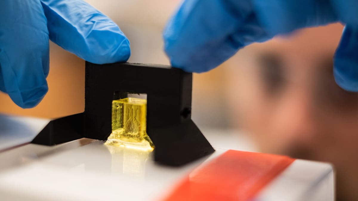
The breakthrough is that the researchers can now, in just a matter of minutes, create soft, water-based, biocompatible gels using a liquid pre-hydrogel solution with intricate internal architecture.
Using this process, the researchers developed a proof-of-concept in the form of a hydrogel lung-mimicking air sac in which airways deliver oxygen to blood vessels. The team also managed to implant bioprinted constructs containing liver cells into mice.
The researchers first 3D printed tissues, then loaded them with primary liver cells before implanting them into mice with chronic liver injury. Amazingly, the liver cells survived the implantation.
Now, the researchers are working on commercializing aspects of their work through Volumetric, a Houston-based startup which designs and manufactures bioprinters and bioinks. However, all of the source data and 3D printable files needed to build the SLATE apparatus is freely available.
“Making the hydrogel design files available will allow others to explore our efforts here, even if they utilize some future 3D printing technology that doesn’t exist today,” says Miller. “We are only at the beginning of our exploration of the architectures found in the human body… We still have so much more to learn.”
Source: Rice University
License: The text of "Researchers 3D Print Tissues with Entangled Vascular Networks" by All3DP is licensed under a Creative Commons Attribution 4.0 International License.