Multicolored 3D Printed Livers Help Doctors Prepare for Tumor Surgery

A medical student in Poland is helping doctors prepare for surgery by creating patient-specific multicolored 3D printed liver models.
Removing a tumor from a liver is no small task. It has always been tricky to prepare for this form of surgery because it is difficult to know the size of a tumor and how close it is to the surrounding vessels.
A medical student at the Jagiellonian University Medical College in Krakow, Poland, has been working on a better way to prepare doctors for surgery. Jan Witowski has been 3D printing model livers to help surgeons see every possible outcome.
“We make those models primarily for complex laparoscopic resections, which aren’t everyday procedures,” explains Witowski. “Those require extra visualization, since it’s important to reduce possible complications and blood loss during the procedure. Surgeons can’t really see what’s under the tissue at specific point of the surgery, and they can’t touch a liver to feel the tumor, because they use laparoscopic approach. They can look at the model during the surgery and realize how far away they are from large vessels, how much more of tumor there is to resect, etc.”
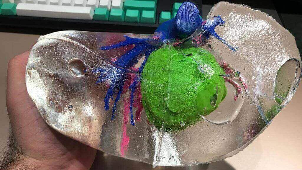
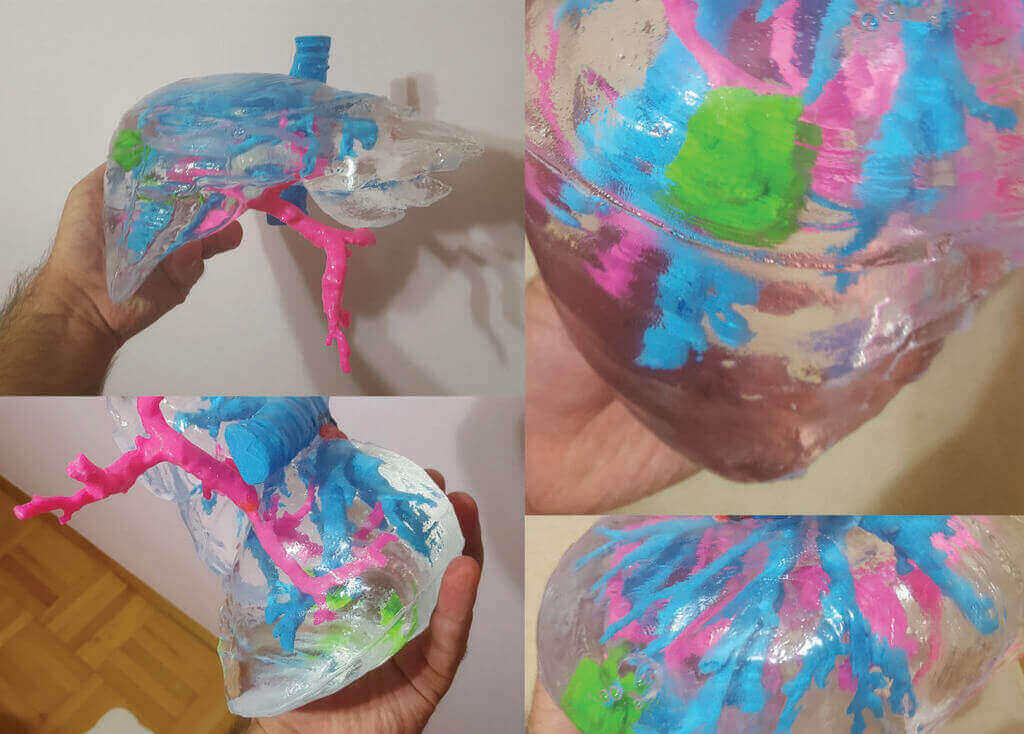
The liver models can be 3D printed with all of the circulatory systems on display. These show up in different colors giving the models a jazzy look. As well as this, Witowski 3D prints any potential issues that might occur during the surgery.
The Making of a 3D Printed Liver Model
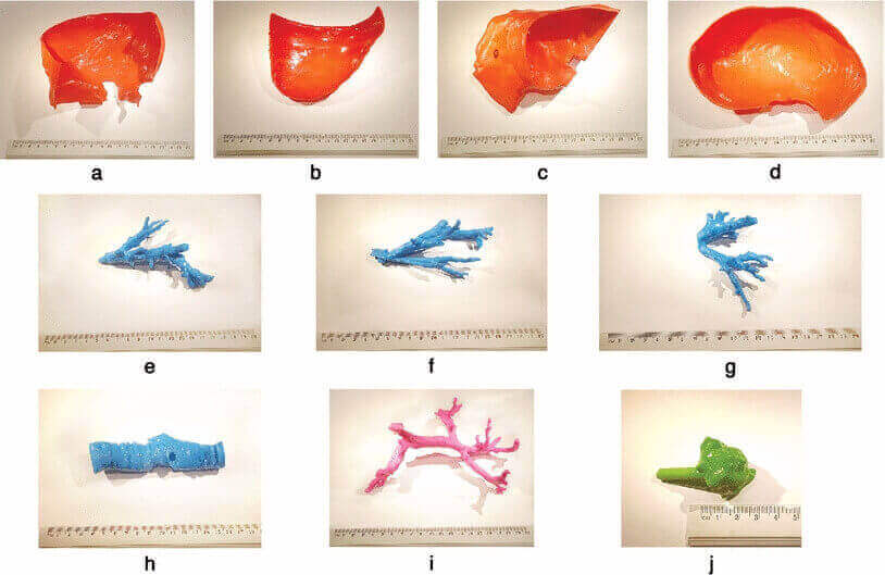
To create the 3D printed livers, Witowski uses CT scans of the patients requiring surgery. By doing this, he can extract stereolithography lithography model from the CRT data.
This model is 3D printed in PLA filament. After this, he builds a “liver parenchyma scaffold.” Into this, he pours silicone material – this is how the surgeon can view the tumors or problems within the liver. You can read more about this process in the paper written by Witowski.
By being able to hold a 3D printed model in their hand, a surgeon understands the shape, weight, and position of the tumor or potential problems. Thanks to 3D printing, this solution is also cost-effective.
Witowski explains: “3D printed models offer great visualization of patient’s anatomy spatial relationships. It is easier to realize the size of a tumor and its proximity to surrounding vessels and liver margins. Another aspect is the possibility to ‘feel the model’ – being table to touch it takes the experience of planning the surgery to another level and makes physical models more realistic than traditional virtually rendered visualizations.”
Source: Tech Crunch
License: The text of "Multicolored 3D Printed Livers Help Doctors Prepare for Tumor Surgery" by All3DP is licensed under a Creative Commons Attribution 4.0 International License.