University of Oxford Develops Method of 3D Printing Living Tissues

An interdisciplinary team of scientists from the University of Oxford and University of Bristol has been able to 3D print cells into living tissue constructs.
A team of researchers from the University of Oxford and the University of Bristol has developed a new method to form living structures with the help of 3D printing.
That’s not all, their method includes using 3D printed laboratory-grown cells. The developments could actually have huge benefits for regenerative medicine and transplantation medicine.
In fact, the method may someday remove the use of animal testing. Also, by developing complex tissues and cartilage, the scientists believe it would be possible to support or repair damaged areas of the human body.
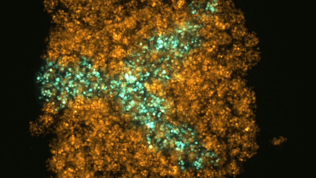
3D printing living tissues turned out to be a complicated process. However, amazingly the results the scientists came up cellular constructs which can mimic, or even enhance, natural tissues.
Lead author and 3D Bioprinting Scientist at Oxford Synthetic Biology is Dr. Alexander Graham. He explains:
‘We were aiming to fabricate three-dimensional living tissues that could display the basic behaviours and physiology found in natural organisms. To date, there are limited examples of printed tissues, which have the complex cellular architecture of native tissues. Hence, we focused on designing a high-resolution cell printing platform, from relatively inexpensive components, that could be used to reproducibly produce artificial tissues with appropriate complexity from a range of cells including stem cells’.
Dr. Adam Perriman from the University of Bristol’s School of Cellular and Molecular Medicine added:
‘The bioprinting approach developed with Oxford University is very exciting, as the cellular constructs can be printed efficiently at extremely high resolution with very little waste. The ability to 3D print with adult stem cells and still have them differentiate was remarkable, and really shows the potential of this new methodology to impact regenerative medicine globally.”
Regenerative Medicine Could Benefit from 3D Printing
Initially, the scientists found that, after printing, the cells would move within the structures and collapse. However, Professor Hagan Bayley of Chemical Biology led a team to create a new way to produce tissues.
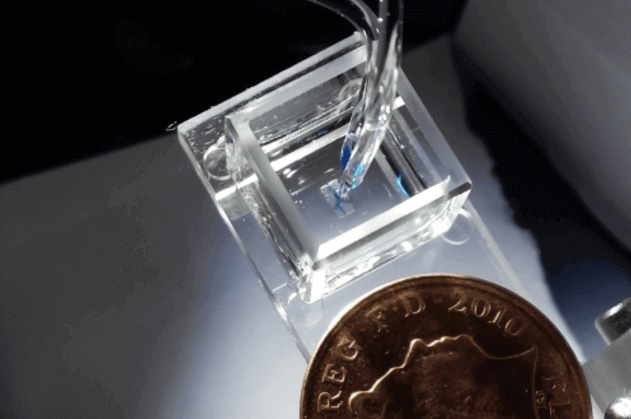
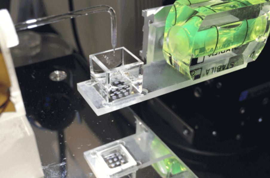
The team created self-contained cells which support the structures and enable them to keep their shape. To do this, they used protective nano liter droplets wrapped in a lipid coating.
Finally, they would 3D print these into living structures. This method improves the survival rate for each individual cell. As well as this, 3D printing allowed the team to build on their current methods. By using 3D printing technology, they could create each tissue one drop at a time. This enables a more favorable resolution.
The hope is that with more work, these materials could hugely improve healthcare worldwide. Dr. Sam Olof, Chief Technology Officer at OxSyBio, explains:
“There are many potential applications for bioprinting and we believe it will be possible to create personalised treatments by using cells sourced from patients to mimic or enhance natural tissue function. In the future, 3D bio-printed tissues maybe also be used for diagnostic applications – for example, for drug or toxin screening.”
Want to find out more? The research was published in the journal Scientific Reports.
Source: University of Oxford News
License: The text of "University of Oxford Develops Method of 3D Printing Living Tissues" by All3DP is licensed under a Creative Commons Attribution 4.0 International License.